
Fig. 2.
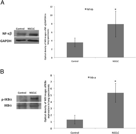
NF − κβ and p-IKBα are significantly more expressed in NSCLC than in control lung tissues. a One representative western blotting image and graphical representation of pixel quantization of NF − κβ and relative total GAPDH of 8 lung tissue specimens. b One representative western blotting image and graphical representation of pixel quantization of p-IKBα and total IKBα in 8 lung tissue specimens. Each experiment was performed three times in duplicate.* = p < 0.05 by t-test analysis. For other details see materials and methods