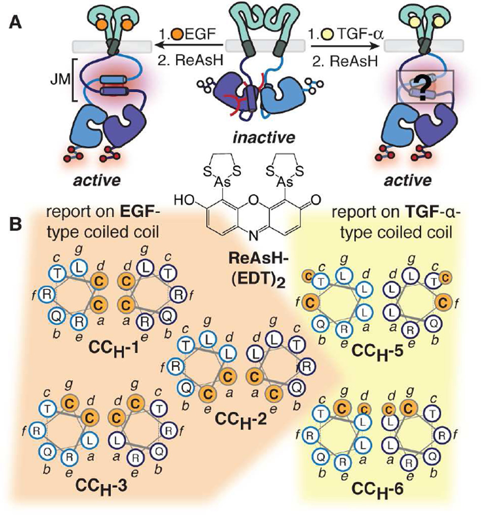
Figure 1.
Probing juxtamembrane segment (JM) structure within full length EGFR on the cell surface using bipartite tetracysteine display (Luedtke et al., 2007; Scheck and Schepartz, 2011) and TIRF microscopy.
(A) EGF and TGF-α induce different structures within the EGFR JM (Luedtke et al., 2007; Scheck et al., 2012; Scheck and Schepartz, 2011).
(B) Helical wheel diagrams of five EGFR variants used previously to distinguish anti-parallel coiled coil arrangements. For sequences, see Figure S1A. In CCH-6, the Cys-Cys motifs are separated axially by a helical turn and do not assemble a ReAsH binding site (see also Figure S1).

