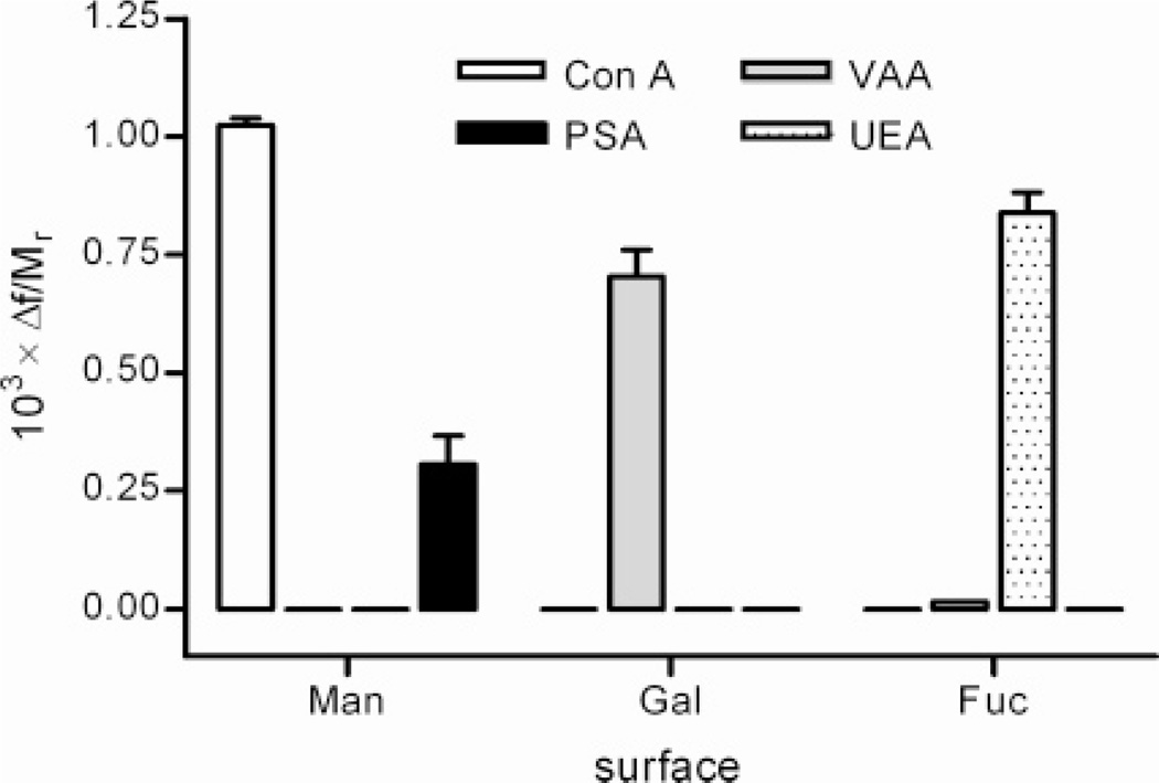
Figure 3.
Determination of binding selectivity. Residual binding (columns) for all four different lectins (Con A, PSA, VAA, UEA) recorded for all three different carbohydrate-derivatized surfaces: α-d-mannopyranoside (Man), β-d-galactopyranoside (Gal), and α-l-fucopyranoside (Fuc). A horizontal bar indicates no detectable residual binding. Protein concentration 10 µM (Con A, PSA, UEA) or 5 µM (VAA).

