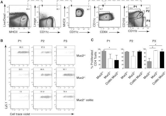
Fig 5. Colitis influences the ability of intestinal DC subsets to induce CD4+ T cell proliferation.
P1, P2 and P3 cells were sorted from pooled MLN from Muc2+/-, Muc2-/- or colitic Muc2-/- and analyzed by flow cytometry. Sorted cells were pulsed with OVA(323–339) peptide prior to co-incubation with CTV-labeled OT-II cells for 5 days. (A) The gating strategy used to identify P1, P2, and P3 populations from MLN cell suspensions is shown. (B) Dot plots show CTV dilution of OT-II cells co-cultured with the indicated iMP subset pulsed with OVA(323–339) peptide. (C) Bar graphs show the frequency of proliferating OT-II T cells ± SD from (B).

