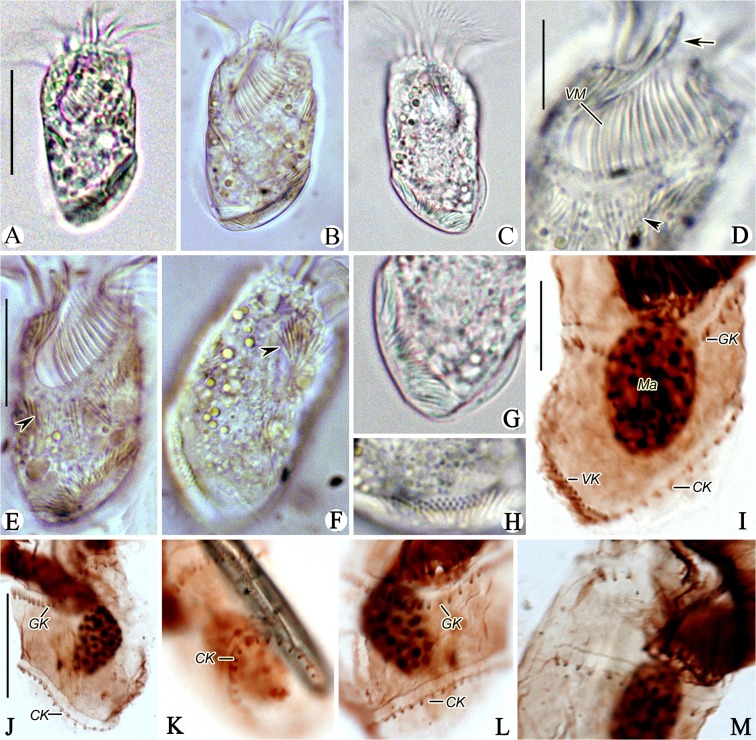
Fig 5. Photomicrographs of Antestrombidium agathae gen. nov., sp. nov. from life (A-H) and after staining with protargol (I-M).
A, B. Ventral views of two individuals; C. Dorsal view; D. Ventral view of anterior part of cell, arrow marks the apical protrusion and arrowhead indicates the extrusomes; E, F. Ventral (E) and Dorsal (F) views showing the distribution of extrusomes (arrowheads); G, H. Posterior portion of cell showing the distribution of extrusomes; I, J. Ventral and dorsal views of posterior portion of cell, showing the somatic kineties; K, L. Left and ventral views showing the GK and CK; M. Right lateral view showing the GK. Legend: CK-circular kinety; GK-girdle kinety; Ma-macronucleus; VK-ventral kinety; VM-ventral membranelles. Scale bars: A-C. 30 μm; D, G-I, L, M. 10 μm; E, F, J, K. 20 μm.

