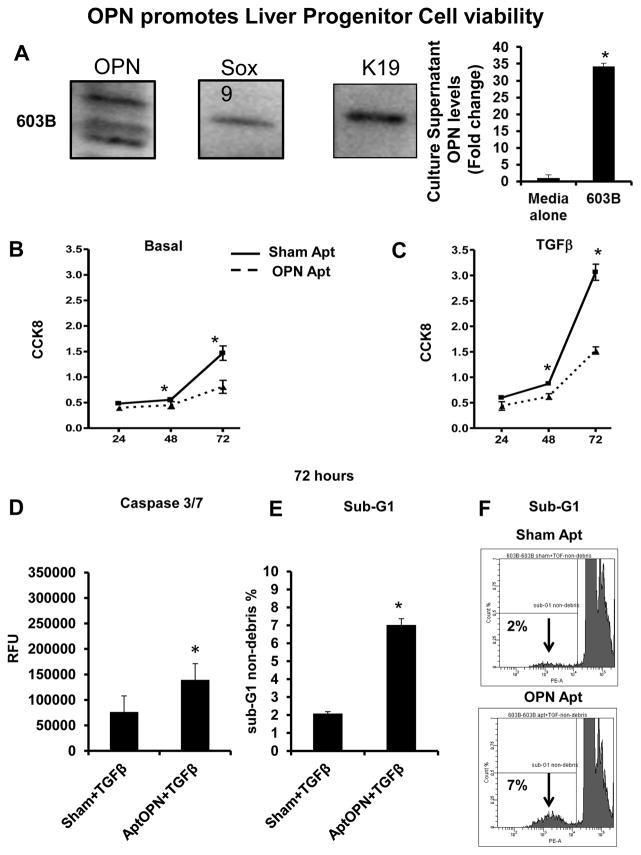
Figure 2. OPN promotes 603B-LPC viability.
603B-LPC were analyzed for OPN, Sox9 and K19 by western blot, and 603B-conditioned medium (CM) assayed using OPN ELISA. In separate experiments, LPC were treated with sham or OPN-aptamers, in the presence or absence of TGF-β. Viability/proliferation was assessed using the CCK8 assay, and apoptotic activity evaluated by caspase-3/7 activity and by sub-G1 analysis (A) OPN, Sox9, and K19 protein expression; OPN levels in 603B-CM. (B) Cell numbers under basal (non-TGF-β) conditions; 24–72 h. (C) Cell numbers under TGF-β conditions; 24–72 h (solid line: sham-aptamer-treated; dashed line: OPN-aptamer-treated). Mean ± SEM (O.D) are graphed. (D) Caspase-3/7 under TGF-β conditions; 72 h. Mean ± SEM (RFU) are graphed. (E) Sub-G1 analysis under TGF-β conditions; 72 h. % of cells in sub-G1 are graphed (F) Representative sub-G1 histograms from sham or OPN-aptamer treated 603B at 72 h. All experiments were performed in triplicate. *p<0.05 vs. respective baseline

