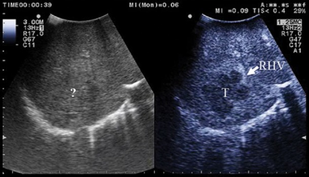
Figure 2.

(a) the margins of the lesion invading the right hepatic vein (RHV) are unclear (?) at intra-operative ultrasound (IOUS); (b) at contrast-enhanced (CE)-IOUS the lesion (T) becomes clearly visible by its margins and its relation with the right hepatic vein (RHV)
