
Figure 5.
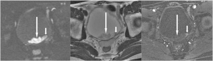
A 41-year-old man presented with gross hematuria due to bladder cancer. Cystoscopy showed a bladder mass. Axial sections of DWI (left) and T2WI (middle) reveal a hyperintense lobulated contour mass (long arrows) in the depending portion of the bladder; and DCE (right) reveals a small early enhanced nodule within this mass, indicating that the tumor (short arrows) is hidden within the blood clot (long arrows).