
Figure 11.
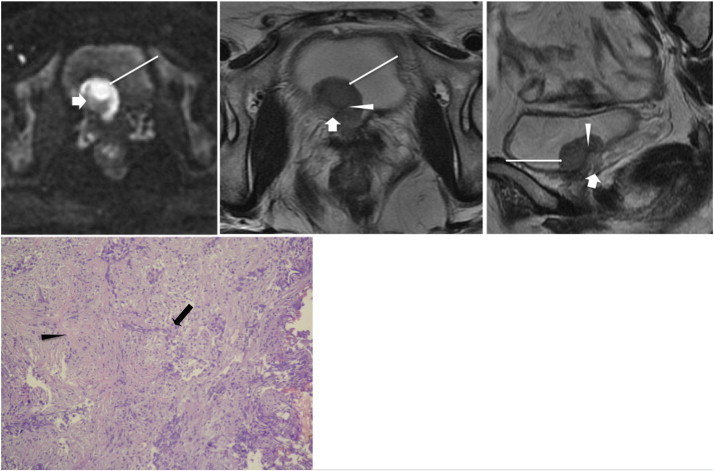
A 53-year-old woman had gross hematuria due to bladder cancer. Axial sections of DWI (top left) and T2WI (top middle) and sagittal section of T2WI (top right) reveal a broad-base tumor (long white arrows) located over the right bladder neck and right bladder base. In addition to a hyperintense tumor area found on DWI, a crescent moon–shaped hypointense area within the central base region is also shown on DWI; and it shows high SI on T2WI (short white arrows), which mimics a thickened submucosa. Pathology (bottom) shows that the tumor (black arrow) infiltrates the muscle layer (black arrowhead) without extending into the perivesical fat, indicating stage T2. Retrospective review of the DWI and T2WI shows that a thin, curvilinear low SI (white arrowheads) could be found between the base of the tumor and the crescent moon–shaped lesion on T2WI, corresponding to the muscle layer; the signal of the muscle layer on DWI could not be identified in the tumor region. The crescent moon–shaped lesion is perivesical fat attracted into the tumor implanted region, not the thickened submucosa.