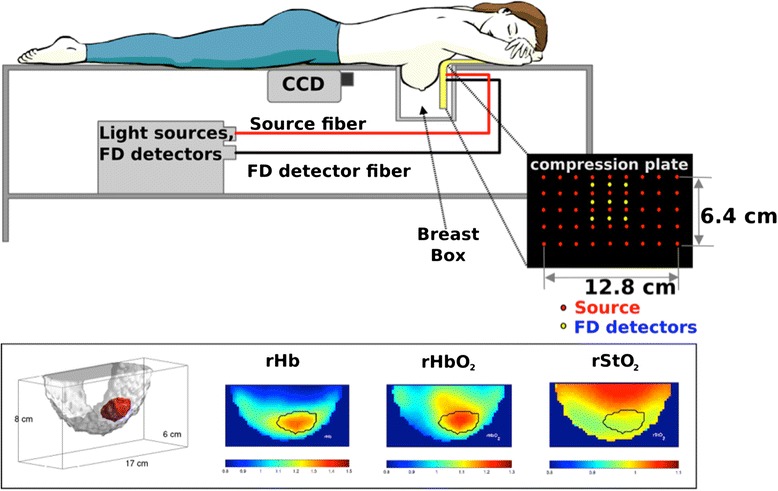
Fig. 1.

Instrumental setup for the diffuse optical tomography (DOT) system, and DOT images acquired from a 53-year-old woman with a 2.2-cm (longest dimension) invasive ductal carcinoma. Bottom left depicts a three-dimensional tumor region (red). The images of rHb, rHbO 2 and rStO 2 are for relative (i.e., tumor-to-normal ratio) deoxyhemoglobin and oxyhemoglobin concentration and tissue oxygenation, respectively. Black solid line in the images indicates the region identified as tumor. FD frequency domain, CCD charge-coupled-device camera
