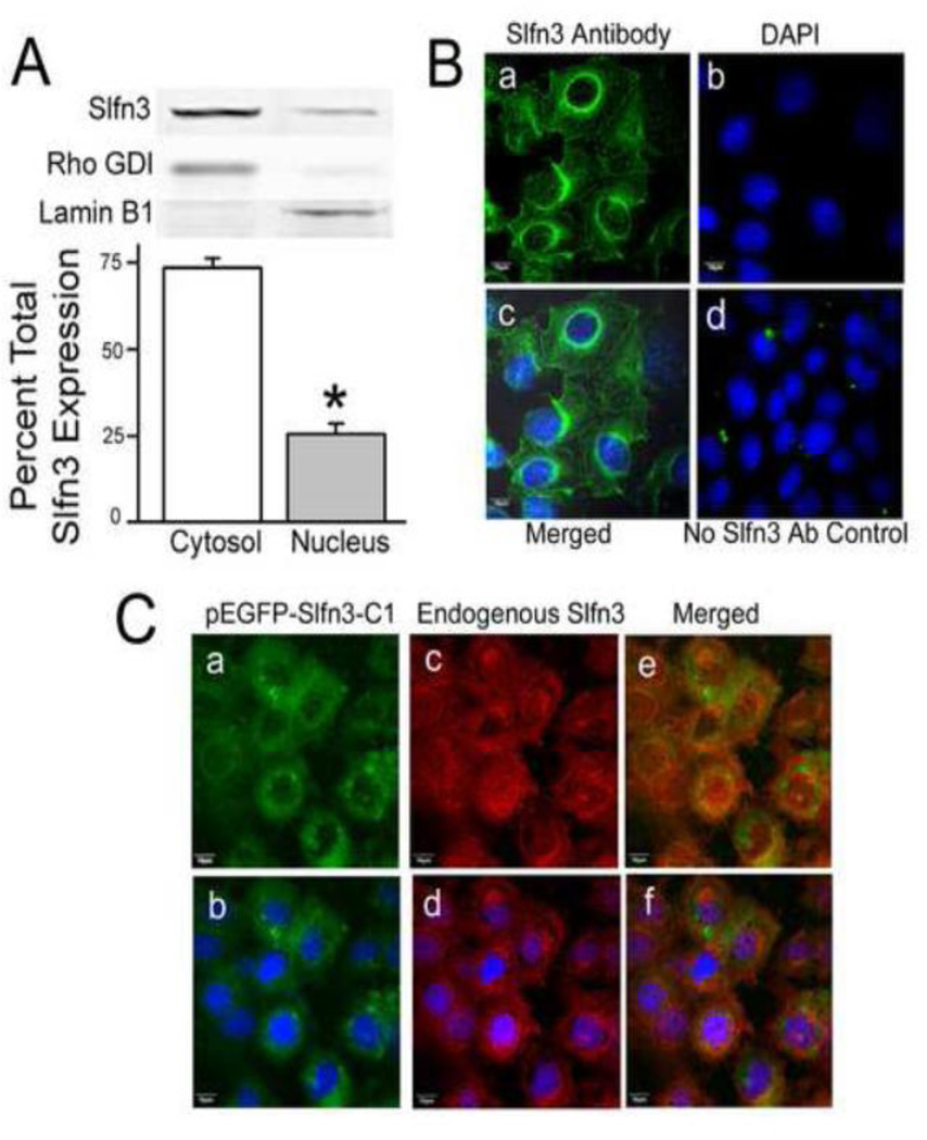
Figure 1. Predominantly cytosolic and modest nuclear localization of exogenously expressed Slfn3 mimics that of the endogenous molecule in IEC-6 cells.
(A) Slfn3 protein abundance is greatest in the cytosol in fractionated rat IEC-6. Bars represent densitometric analysis as a ratio of the total immunoreactivity (*p<0.05, n=8). A representative gel is shown above; Rho GDI and Lamin B were used as cytosolic and nuclear markers. (B) Confocal images confirm that endogenous immunoreactive Slfn3 is found in the nucleus but is predominantly localized to the cytosol: (a) Slfn3 primary antibody/FITC secondary antibody, (b) DAPI nuclear stain, (c) merged image, and (d) no primary antibody control with DAPI stain. Representative of 8 similar images. (C) (a) Exogenously expressed Slfn3 (pEGFP-Slfn3-C1) and (b) endogenous Slfn3 co-localize in IEC-6 cell (c). Cytosolic/nuclear expression is further delineated with nuclear DAPI staining (b, d, f). Representative of 6 similar.

