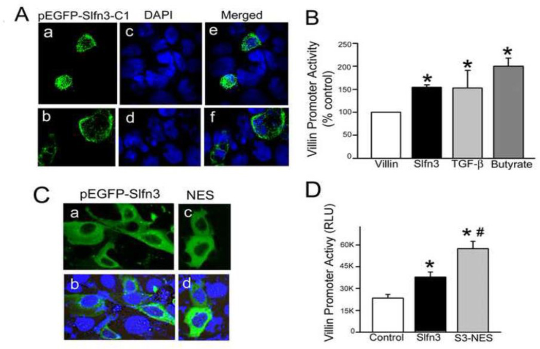
Figure 2. Exogenously expressed Slfn3 stimulates villin promoter activity from a cytosolic location in Slfn3-null Caco-2 cells.
(A) Two representative confocal micrographs of pEGFP-Slfn3-C1 expressed in Caco-2 cells demonstrate strong cytosolic and minor nuclear expression (a, b). DAPI-stained nuclei reflect the number of non-transfected cells (c, d), Merged images (e, f) confirm that the pEGFP-Slfn3-C1 construct is expressed in Caco-2 as it is in IEC-6 cells. (B) Exogenous Slfn3 enhances villin promoter activity similarly to TGF-β and sodium butyrate (*p<0.05; n=3–6). (C) Cellular localization of GFP-tagged wild-type Slfn3 (pEGFP-Slfn3) transfected into Slfn3-null Caco-2 cells (a, b) and cytosolic localization with the addition of a nuclear exclusion sequence (pEGFP-Slfn3-NES; c, d) is shown in these representative confocal micrographs. (D) Villin promoter activity is greater in Caco-2 cells transfected with the Slfn3-NES construct (S3-NES) than with the full-length construct (Slfn3) (*p<0.05 vs control, #p<0.05 vs full length by one way ANOVA, n=4).

