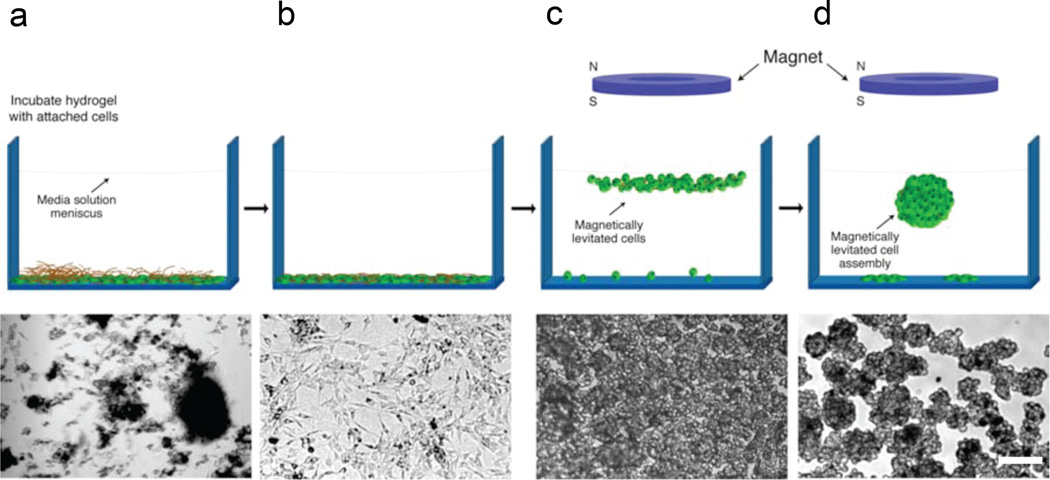
Figure 2. 3D cell culture with magnetic-based levitation.
The top row shows the general cell levitation strategy and the bottom row shows the corresponding optical micrograph of neural stem cells at each stage. a, Hydrogel is dispersed over cells and the mixture is incubated. The dark blotches are fragments of hydrogel. b, Washing steps remove non-interacting hydrogel fragments. Fractions of phage, gold, and MIO nanoparticles enter cells or remain membrane-bound. c, Application of an external magnet causes cells to rise to the air-medium interface. Image shows culture 15 min after levitation. d, After 12 h of levitation, characteristic multicellular structures form (single structure is shown in the schematic). Scale bar, which applies to bottom row, 30 µm.

