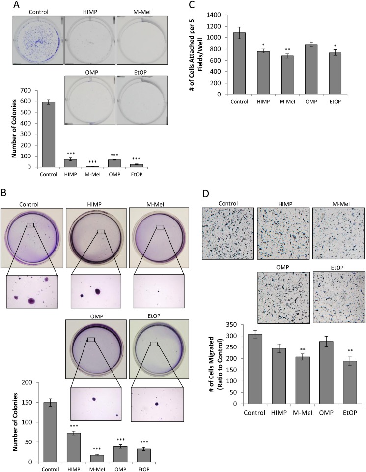
Fig 3. Effects of imidazopyridine derivatives on the tumorigenicity of LNCaP C-81 cells.
(A) Clonogenic assay on plastic wares. LNCaP C-81 cells were plated in six-well plates at densities of 20, 200, and 2,000 cells/well. After 24 hours, attached cells were treated with respective compounds at 10 μM concentrations of imidazopyridine derivatives or solvent alone as control. Cells were fed on days 3, 6, and 9 with fresh culture media containing respective inhibitors. On day 10, cells were stained and the number of colonies counted. The photos of representative colony plates were taken from plates seeded with 2,000cells/well, and the number of colonies shown was counted also from plates seeded with 2,000cells/well. Minimal colony formation was observed at densities of 20 and 200 cells/well. Results presented are mean ± SE; n = 2x3. ***p<0.0001. (B) Anchorage-independent soft agar assay. LNCaP C-81 cells were plated at a density of 5 x 10⁴ cells/35mm dish in 0.25% soft agar plates. The following day, cells in doublets or greater were marked and excluded from the study. Media were added every three days, and at the end of 5 weeks, colonies formed were stained and counted. Representative images of colonies are shown (above) and the colony number was counted (below). The experiments were performed in duplicate with 3 sets of independent experiments. Results presented are mean ± SE; n = 2x3. *** p<0.0001. (C). Cell adhesion assay on plastic wares. Cells were suspended in treatment media for 30 minutes before being plated in 6-well plates at 3 x10³ cells/cm2 using the same treatment media. Cells were allowed to adhere for one hour, fixed and stained by 0.2% crystal violet solution (50:50, water:MeOH). The total number of cells in five fields at 40x magnification for each well was counted. The experiments were performed in triplicate with 3 sets of independent experiments. Results presented are mean ± SE; n = 3x3. *p<0.05; **p<0.01. (D). Cell migration transwell assay. Cell migration was assessed via Boyden chamber. An aliquot of 5 x 10⁴ C-81 cells was seeded in the insert of 24-well plates in media containing 10 μM respective compounds with solvent alone for control in both upper and lower chambers. After 24-hour incubation, the migrated cells were stained and those cells remaining in the upper chamber were removed via cotton swab. Cells which had migrated through to the lower chamber were counted. Representative images are shown at 40x magnification. The experiments were performed in triplicate with 3 sets of independent experiments, and the results presented are mean ± SE; n = 3x3, *p<0.05; **p<0.005.

