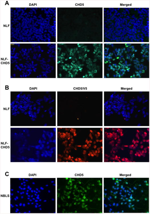
Figure 2. Nuclear localization of CHD5-IF.

(A) Parental NLF cells (top row) and NLF–CHD5 (second row) were cultured for 48 h. Cells were immunostained with CHD5 as indicated. Nuclei were stained with DAPI (left panels). Nuclear localization of CHD5 was observed when cells overexpressed CHD5 (middle panels), whereas no nuclear staining was observed in control NLF cells (upper middle panel). Merged images are shown on right. (B) IF images indicating nuclear expression with tagged V5–His antibody. (C) Nuclear expression of CHD5 in NBLS cells expressing endogenous CHD5.
