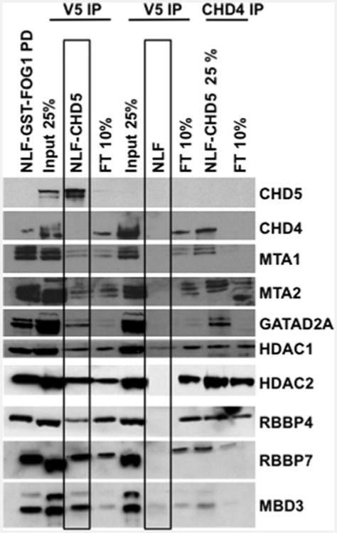
Figure 4. Detection of CHD5–NuRD components: IP and Western analysis.

Nuclear extracts from NLF–CHD5 and NLF were immunoprecipitated as indicated. Subsequent proteins from IP and GST–FOG1 pull-down (left lane) were subjected to 4%–12% PAGE. Extracts from NLF following GST–FOG1 pull-down served as negative control. Individual NuRD components were detected by Western blotting with antibodies specific to each NuRD component. The boxes around the NLF–CHD5 and NLF lanes highlight the identification of all canonical NuRD components in NLF–CHD5 and not in NLF as a negative control, as these cells do not express CHD5 endogenously. FT, flow through.
