
Fig. 4.
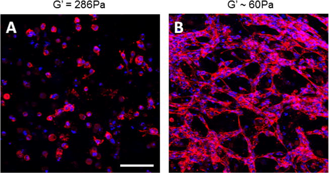
(A) Endothelial cells do not elongate or form MVN in 1 mg ml−1 KFE gels, which do not contain integrin binding sites, but (B) do form MVN in 1 mg ml−1 KFE-RGD gels, which do contain integrin binding sites. Cells are stained with Alexa 633 phalloidin (red), and nuclei are stained with DAPI (blue) to highlight networks and allow for quantification. Stiffness of (A) was measured by rheology, while (B) was calculated using Eq. (1). Scale bar is 100 μm.