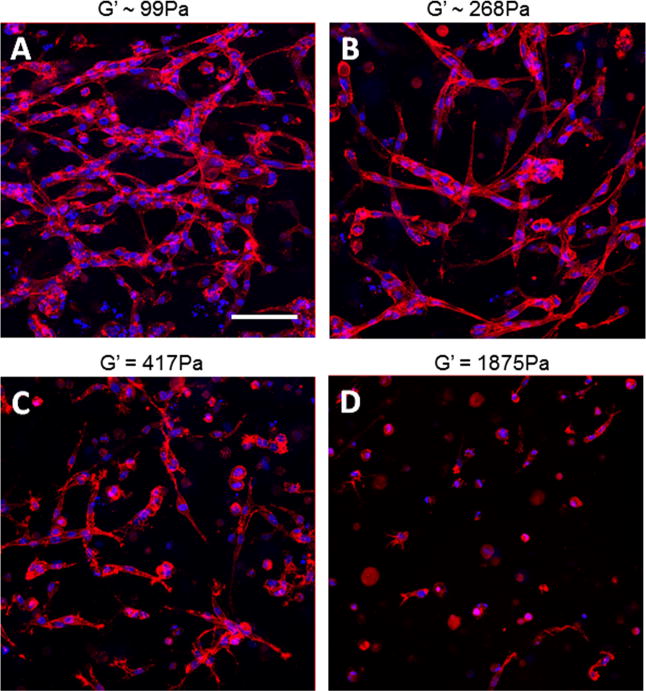
Fig. 5.

(A–D) Images of phalloidin (red) and DAPI (blue) stained HUVEC showing decreased elongation and MVN formation of endothelial cells as stiffness is increased by increasing KFE, while binding site density and thus KFE-RGD are held constant. Holding KFE-RGD concentration constant at 1 mg ml−1, (A) 0.1, (B) 0.5 (B), (C) 1 or (D) 2.5 mg ml−1 KFE was added to increase stiffness. Stiffnesses of (A) and (B) were estimated using Eq. (1), while (C) and (D) were measured by rheology. Scale bar is 100 μm.
