Fig. 1.

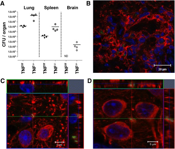
Dissemination of M. tuberculosis to the brain in TNF−/− mice. a TNFf/f and TNF−/− mice were infected by aerosol inhalation at a dose of 200–500 CFUs/lung of M. tuberculosis, and mycobacterial burden in brains, lungs and spleens was determined at day 33 post-infection. b Foci of disseminated fluorescent bacilli (green) surrounded by CD11b+ microglia/macrophages (red) were present in the TNF−/− brain parenchyma. c, d The fluorescent image of intracellular M. tuberculosis GFP-expressing bacilli (green) found in the neurons (red) of TNF−/− mouse, labelled with c MAP2 and d β-III-tubulin. The orthogonal projection of confocal Z-stacks confirmed the cytosolic location of the bacilli in the x–y plane. All nuclei were labelled with DAPI (blue in b–d). Scale bars: b 20 μm; c, d 5 μm. The data represents two independent experiments
