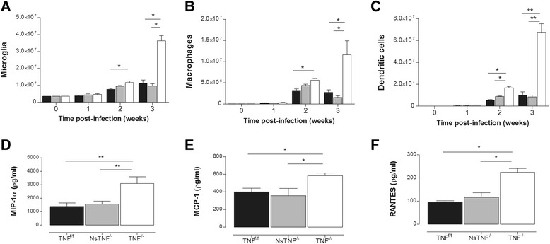
Fig. 6.

TNF regulates innate immune cell proliferation and recruitment during CNS-TB. TNFf/f (black), NsTNF−/− (grey) and TNF−/− (clear) mice were infected with M. tuberculosis H37Rv at a dose of 1 × 10³–1 × 10⁴ CFUs/brain by intracerebral inoculation. The number of a microglia, b macrophages and c dendritic cells was analysed by flow cytometry at 0, 1, 2 and 3 weeks in infected brains. The chemokines d MIP-1α, e MCP-1 and f RANTES were measured by ELISA in the brains of infected mice at 3 weeks post-intracerebral infection. Data (mean ± SEM of the CFUs of 5–6 mice per time point) are representative of repeat experiments. *p ≤ 0.05; **p ≤ 0.01
