
Fig. 3.
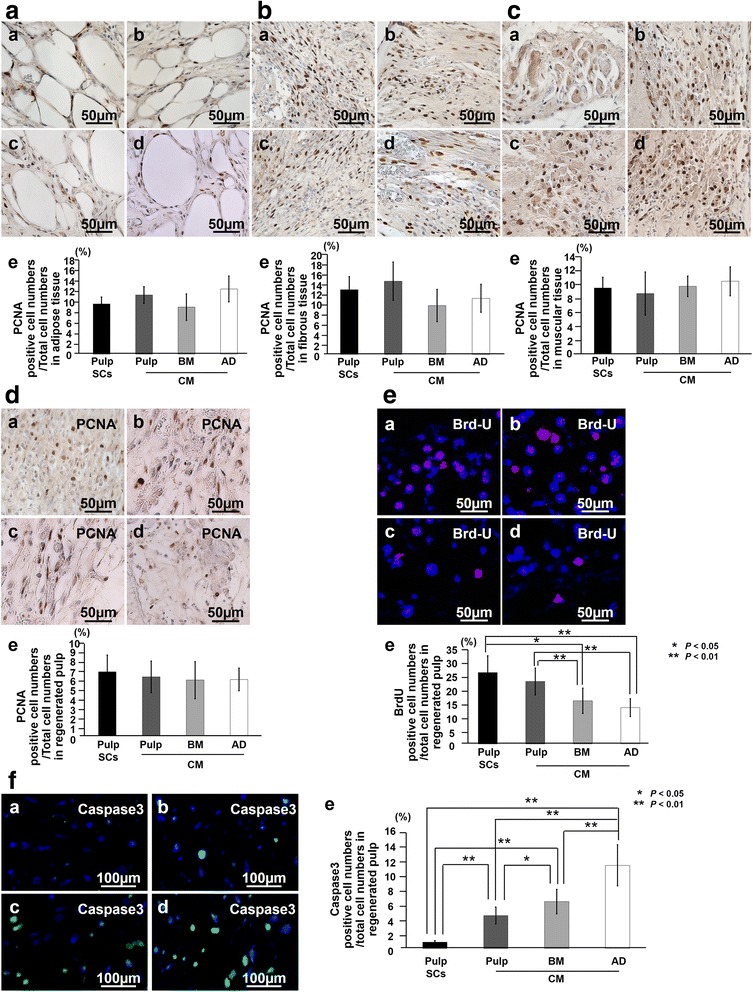
The effect of conditioned medium (CM) on proliferation, migration, and anti-apoptosis in the surrounding tissues of the transplanted teeth and the regenerated pulp. Immunostaining with proliferating cell nuclear antigen (PCNA) in adipose (AD) tissue (a), fibrous tissue (b), muscle tissue (c), and regenerated pulp tissue (d) 7 days after transplantation. (a) Pulp CD31− side population (SP) cells, (b) CM from pulp CD31− SP cells, (c) CM from bone marrow (BM) CD31− SP cells, and (d) CM from AD of CD31− SP cells. (e) Ratio of PCNA-positive cell numbers to the total cell numbers in the each tissue. e Bromodeoxyuridine (BrdU) (red) merged with Hoechst 33342 (Blue) in the regenerated pulp tissue on day 7. (e) Ratio of BrdU-positive cell numbers to the total cell numbers. f Immunostaining with caspase 3 (green) merged with Hoechst 33342 (Blue). (e) Ratio of caspase 3-positive cell numbers to the total cell numbers of regenerated pulp. Seven days after transplantation of (a) pulp CD31− SP cells, (b) CM from pulp CD31− SP cells, (c) CM from BM CD31− SP cells, and (d) CM from AD of CD31− SP cells. Data are expressed as mean ± standard deviation of four determinations. *P < 0.05, **P < 0.01