
Fig. 7.
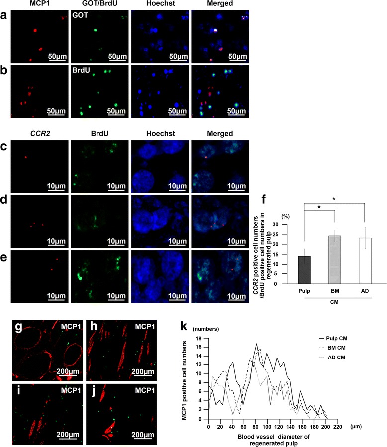
Localization of monocyte chemotactic protein 1 (MCP1) and its receptor, CCR2, related to the transplanted cells and the endogenous migrated cells in the regenerated pulp. a, b Immunostaining of MCP1 (red) merged with glutamic-oxaloacetic transaminase 2 (GOT2) or bromodeoxyuridine (BrdU) (green) and Hoechst 33342 (blue) 7 days after transplantation of pulp CD31− side population (SP) cells. c-e In situ hybridization and immunohistochemical analysis of expression of CCR2 (red) merged with BrdU (green) 7 days after transplantation of conditioned media (CM) from pulp (c), bone marrow (d), and adipose CD31− SP cells (e). f Ratio of CCR2-positive cell numbers to BrdU-positive cell numbers in the regenerated pulp. Data are expressed as mean ± standard deviation of four determinations. *P < 0.05. g-j MCP1-positive cells (green) in proximity to the newly formed vessels immunostained with rat endothelial cell antigen 1 (RECA1) (red) 28 days after transplantation of pulp CD31− SP cells (g), CM of pulp (h), CM of bone marrow (i), and CM of adipose CD31− SP cells (j). Note no co-localization. k The number of MCP1-positive cells related to the diameter of blood vessels