
Figure 7.
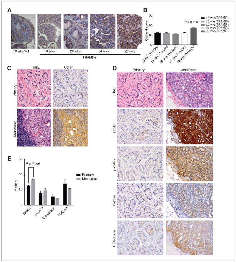
Cofilin overexpression correlates with prostate cancer progression to metastasis A and B, cofilin profiling in TRAMP mouse model. TRAMP transgenic mice develop prostate adenocarcinoma with increasing age, resembling progression of human prostate cancer to metastasis. Prostate sections of increasing grade and metastatic tumors (16–28 weeks) were profiled by immunostaining for cofilin expression; WT mouse prostate tissue (16 weeks) was used as control; magnification, ×40. Quantitative evaluation of CFL immunoreactivity, as determined by the H-scoring, shows a significant increase in metastatic tumors from 28-week-old TRAMP mice (P = 0.001) compared with early-stage tumors. C–E, cofilin expression profile in human prostate cancer. C, hematoxylin and eosin staining and CFL immunostaining in serial sections of prostate tumors; characteristic image of a metastatic lesion to lymph nodes exhibiting intense cofilin immunoreactivity, compared with the primary tumor from the same patient (absence of CFL expression). Magnification, ×100. D, representative images of immunostaining for cofilin, p-cofilin, E-cadherin, and palladin on primary and metastatic prostate cancer. E, quantitative analysis of protein immunoreactivity (from D). There was a significant increase in cofilin levels in metastatic specimens compared with primary tumors (P = 0.005).