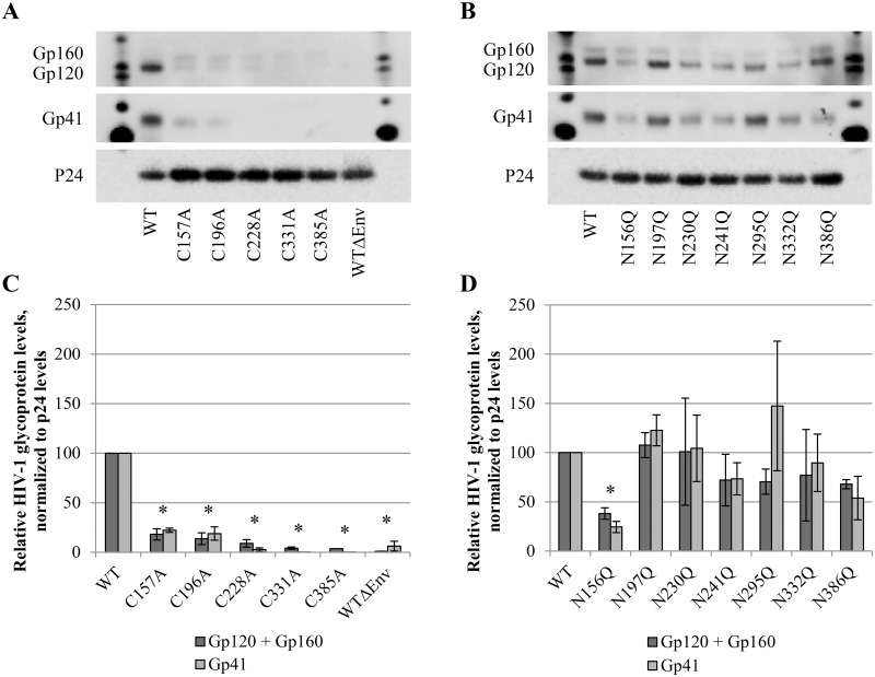
Fig 4. Western blot analysis of envelope glycoprotein incorporation in WT and mutant virus particles.
Virus was concentrated, lysed and subjected to western blotting. Gp120, gp41 and p24 were detected in virus lysates of (A) mutants lacking a disulphide bridge and (B) N-glycosylation site mutants. (C-D) Quantification of protein levels was performed using the BioRad Image Lab Software, based on panels A and B. Gp160, gp120 and gp41 levels are presented after normalization to the p24 levels. Graphs represent the mean ± SEM based on 2–4 independent experiments. The difference between WT and mutant virus was considered to be significant when the p value calculated using the student’s t-test was <0.05 (* = p<0.05, for both gp120+gp160 and gp41).

