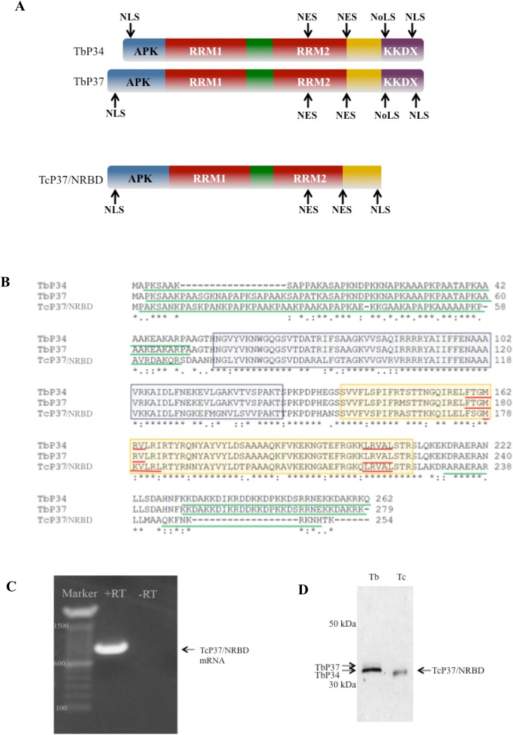
Fig 1. TcP37/NRBD is a homolog of TbP34 and TbP37.
Panel A. Schematic diagram of TbP34, TbP37, and TcP37/NRBD. APK: alanine-proline-lysine rich region. RRM: RNA Recognition Motif. KKDX: Region containing repeats of lysine, lysine, aspartic acid, and X representing any amino acid. NES: Nuclear Export Signal. NLS: Nuclear Localization Signal. NoLS: Nucleolar Localization Signal. Panel B. Multiple sequence alignment of TbP34, TbP37 and TcP37/NRBD protein. An asterisk indicates identity, a colon indicates conserved substitution, and a period indicates semi-conserved substitution. The amino acids of the proteins are indicated by terminal numbers. Blue and yellow boxes indicate RRM1 and RRM2 domains respectively, solid red underlines denote the predicted nuclear export signal, and solid green underlines indicate predicted nuclear localization signals of these proteins. Panel C. TcP37/NRBD is endogenously expressed as mature mRNA in epimastigotes. RT-PCR was performed on T. cruzi total RNA. RT: control with no reverse transcriptase, +RT: amplified specific product, Marker: DNA molecular weight markers (bp). Panel D. Western blot analysis of 2 micrograms of whole cell extract protein prepared from T brucei procyclic cells (Tb) and T. cruzi epimastigote cells (Tc). The membrane was probed using the P34/P37 antibody. Two bands are visible in the Tb lane (TbP34 and TbP37), and a single band is observed for Tc (TcP37/NRBD). Numbers indicate molecular weight markers.

