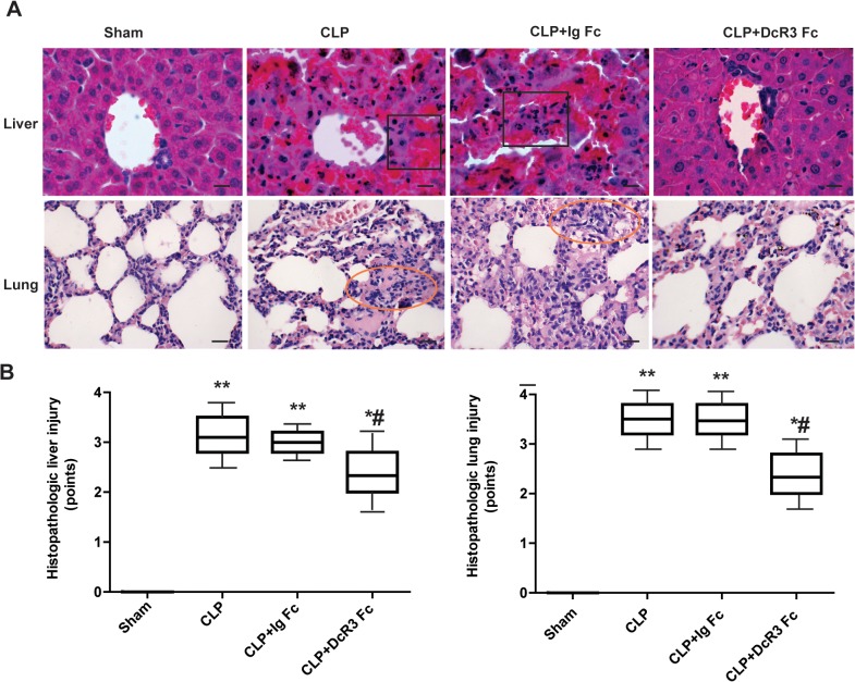
Fig 2. Histopathological changes in septic mice after DcR3 Fc administration.
Liver and lung tissues were harvested 24 h after CLP. Hematoxylin–eosin staining was performed to examine histopathological changes (400 ×). (A) Representative images for each group. Lung injury was characterized by thickening of the alveolar wall, accumulation of neutrophils into the interstitium and impaired alveoli in CLP mice (circle areas). Liver injury was evidenced by swollen hepatocytes, inflammatory infiltration and hemorrhagic necrosis in CLP mice (square areas). DcR3 Fc treatment protected mice against sepsis-induced liver and lung damage. Lung displayed less alveolar destruction and alveolar epithelial hyperplasia when treated with DcR3 Fc. Hepatocytes showed less ballooning lesions. (B) The severity of lung and liver injury was scored as described above. Scale bars in right lower corner represent 25μm. **p <0.01, *p <0.05 vs. sham; #p <0.05 vs. CLP.

