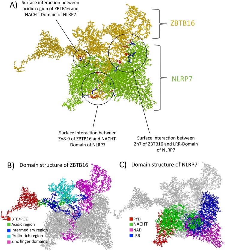
Fig 5. Blind docking of ZBTB16 and NLRP7 threaded protein models.
The figure is split into three panels, each representing the ZBTB16/NLRP7 dock in differently colored formats. The models are depicted in stick form. A. The first panel shows the proteins ZBTB16 and NLRP7 in yellow and green. The interface regions are marked and specified. The red residues on the interface regions belong to ZBTB16, while the blue ones belong to NLRP7. B. and C. Both panels show the same docks but with the individual domains of ZBTB16 and NLRP7, respectively, colored according to the code described below. The apposing protein in each panel is uniformly colored in grey.

