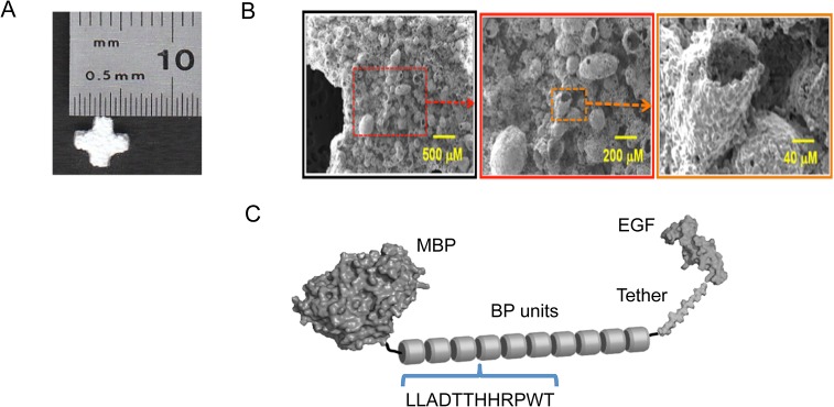
Fig 1. Structures of ßTCP scaffolds and EGF-ßTCP binding peptide (BP-T-EGF) fusion protein.

Macroscopic appearance of a 5mm ßTCP Therilok cross-shaped scaffold used for cell culture experiments (A). These scaffolds were crushed to a coarse powder for phage panning to select binding peptides. SEM micrographs of the surface of ßTCP scaffolds at low, intermediate and high resolution as indicated by scale bars (B), Structure of the fusion protein “BP10-T-EGF” comprising the 12-amino acid ßTCP-binding peptide fused to EGF by flexible protease-resistant tethers flanking a coil domain (C; see S1 Table for specific sequence).
