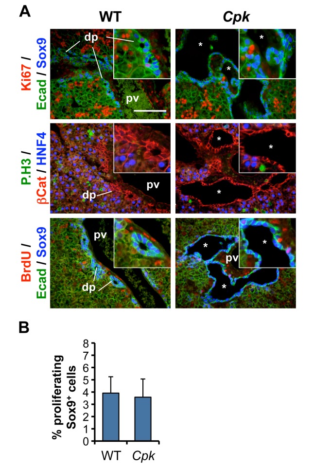
Fig 2. No excessive proliferation during biliary cyst formation in mouse Cpk embryonic livers.
(A) Immunofluorescent staining against the indicated markers was performed on tissue slides from WT and Cpk mouse livers at E17.5. Proliferation rates, revealed by Ki67, P.H3 and BrdU stainings, are low in WT and Cpk cholangiocytes, while the rest of the liver exhibits higher levels of proliferation. *, cysts; pv, portal vein; dp, ductal plate. Bars: 100 μm. Insets: 2-fold higher magnifications. (B) Percentage of proliferating Sox9+ cholangiocyte precursors, calculated using the formula (nKi67+;Sox9+/nSox9+) x 100, like in Fig 1B (n = 3 for each genotype). Data are mean +/- SD. Two-tailed t-test was performed, and a p-value <0.05 was considered significant.

