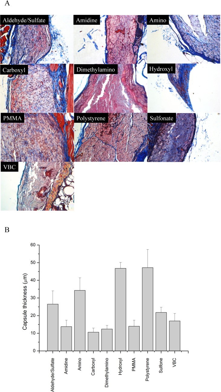
Fig 2. Masson’s Trichrome staining of representative sections subcutaneously injected.
(A) Representative sections stained with Masson’s Trichrome are shown for the various polystyrene particles injected into mice. (Magnification 20×, images are 540 × 360 μm2) (B) Quantification of capsule thickness. Error bars represent the standard deviation.

