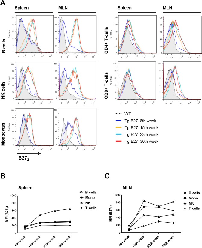
Fig 4. Detection of cell-surface B272 in leukocyte populations of HLA-B27 transgenic rats at different ages.
(A) Representative flow cytometry analysis of cell-surface B272 expression in leukocytes populations of Tg rats at different ages (6, 15, 23 and 30 weeks) from spleen and MLNs. WT leukocytes represent the control population where B272 is absent. (B) MFI values plotted of positive B272 stains from splenocytes. (C) MFI values plotted of positive B272 stains from MLNs. Detection of cell-surface B272 homodimers was performed using HD6-biotinylated and detected by streptavidin-APC. HD6 had been previously assessed as an antibody capable of recognizing cell-surface B272 in human [12] and rat [41] leukocyte populations. Antibody panels: CD4+ T-cells (+CD3, +CD4), CD8+ T-cells (+CD3, +CD8), NK (+CD161a,—lineage), B cells (+CD45RA,-lineage) and Monocytes (+CD172a,—RP-1).

