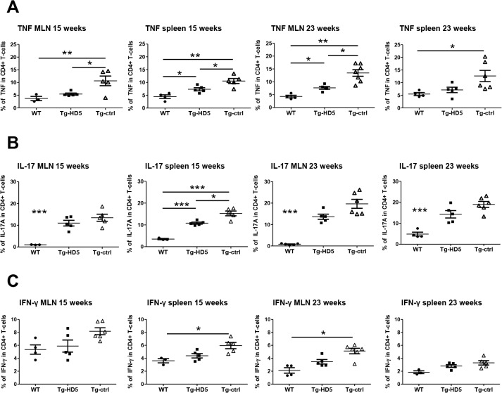
Fig 6. Reduced expansion of pro-inflammatory CD4+ T-cells from spleen and MLN in Tg-HD5 rats.
In vitro stimulated cells obtained from Tg-HD5, Tg-ctrl and WT-littermates were assessed by ICS for the presence of pro-inflammatory cells expressing TNF (A), IL-17 (B) and IFN-γ (C). MLN and spleens cells were obtained at week 15 (n = 5) and at week 23 (n = 5). Results are plotted as the percentage of CD4+ T-cells gated positive for TNF, IL-17 and IFN-γ. Values are expressed as mean±SEM. *p<0.05, **p<0.01, ***p<0.005 as determined by one-way ANOVA followed by Bonferroni post-hoc analysis.

