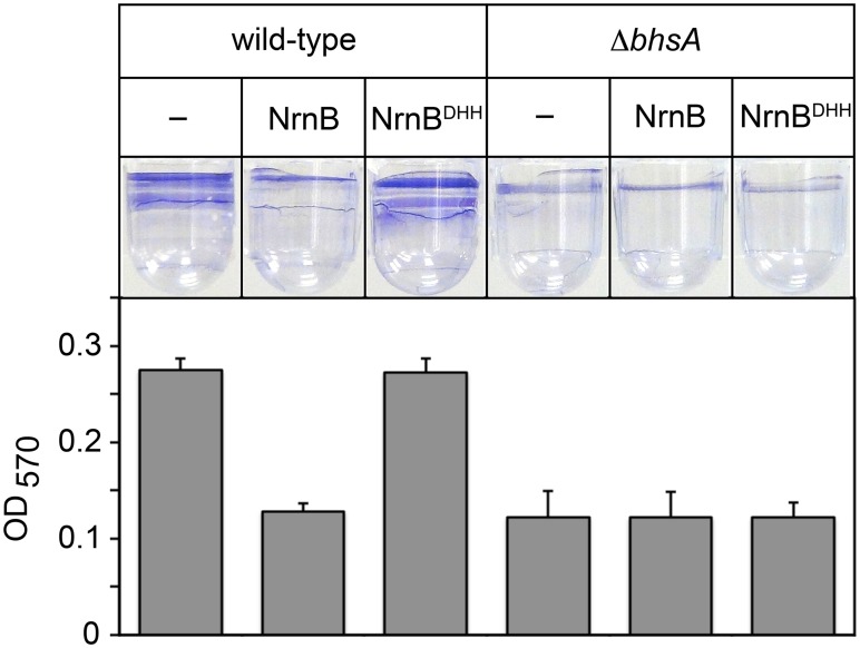
Fig 2. The PDI-dependent increase in bhsA expression contributes to biofilm formation in E. coli.
Top shows images of representative wells containing biofilms stained with crystal violet dye. Graph shows averages +SEM of the amount of crystal violet staining observed from at least four independent measurements. Assays were done using wild-type MG1655 cells or MG1655 cells carrying a deletion of bhsA (ΔbhsA). Cells carried an empty plasmid (–) or a plasmid that directs the synthesis of NrnB or NrnBDHH.

