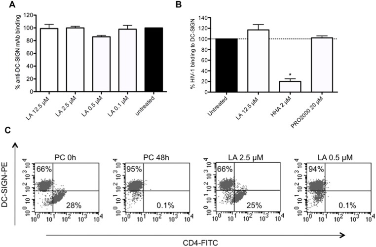
Fig 3. LA inhibits HIV-1 DC-SIGN-related transmission to uninfected CD4+ target T cells.
(A) Raji.DC-SIGN+ cells were pretreated with or without various concentrations of LA for 30 min. The binding of PE-conjugated anti-DC-SIGN mAb (clone DCN46) was measured by flow cytometry. The bars represent the % of anti-DC-SIGN binding relative to the untreated conditions. Mean ± SEM of 3 independent experiments is shown. (B) Effect of LA, HHA and PRO2000 on the capture of HIV-1 strain HE (R5/X4) by DC-SIGN on Raji.DC-SIGN+ cells. HIV-1 virions were pre-incubated for 30 min. with the test compounds before being exposed for 1h to Raji.DC-SIGN+ cells. After washing the cells, p24 HIV-1 Ag ELISA was used to quantify the amount of cell-associated virus. Bars represent the % virus binding relative to untreated conditions. Mean ± SEM of 3 independent experiments is shown with *p<0.05, according to Student’s T-test. (C) HIV-1 HE (R5/X4) virions were exposed to Raji.DC-SIGN+ cells for 1 h. After extensive washing, Raji.DC-SIGN/HE cells were cocultivated with or without compound pretreated uninfected CD4+ target C8166 T cells for 48h. Cocultures were stained with PE-conjugated anti-DC-SIGN and FITC-conjugated anti-CD4. The % of positive gated cells in the quadrants of the dot plots is given. One representative experiment out of 3 is shown.

