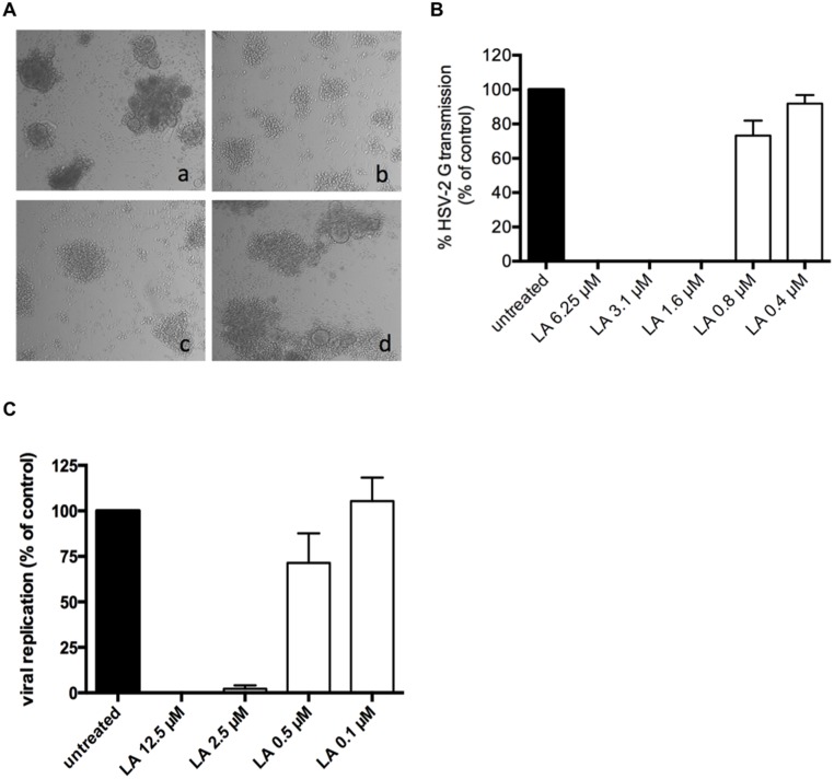
Fig 5. LA inhibits HSV-2 DC-SIGN-related transmission to uninfected target T cells.
(A) DC-SIGN+ cells were generated out of buffy coat and exposed to HSV-2 G for 2 h. After several washing steps, virus exposed cells were cocultivated with uninfected CD4+ MT-4 cells for 5 days. Viral-induced cytopathicity was first scored microscopically. Shown here are the light microscopic pictures of cocultivated HSV-2 G-exposed DC-SIGN+ cells and uninfected MT-4 cells 5 days post-cocultivation (a); the effects on HSV-2 G transmission and replication in the presence of 12.5 μM (b), 2.5 μM (c) and 0.5 μM LA (d). One representative experiment out of 4 individual donors is shown. (B) Bars represent the % HSV-2 G transmission and replication in MT-4 cells after 5 days of cocultivation, relative to the positive control. Dying MT-4 infected cells were detected by flow cytometry side-scatter analyses. Mean ± SEM out of 4 individual donors is shown. (C) MDDCs were generated from buffy coat and exposed to HSV-2 G for 2h. After several washing steps, virus exposed MDDCs were co-cultivated with uninfected CD4+ MT-4 cells for 5 days. Viral-induced cytopathicity was scored light microscopically and supernatant was retained for further analysis of viral content. Bars represent the percentage of viral replication after 5 days of co-cultivation, relative to the positive untreated control. Mean ± SEM of 3 independent experiments is shown.

