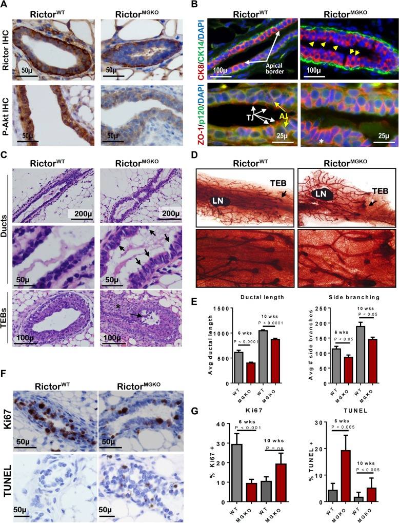
Fig 1. Loss of Rictor disrupts mammary branching morphogenesis in vivo.
A. IHC for Rictor (upper panels) and P-Akt S473 (lower panels) in mammary gland sections from 6 week-old virgin mice. B. IF for CK8, CK14, ZO-1 and P120 in mammary gland sections. Yellow arrows indicate apically mis-localized nuclei in RictorMGKO tissue in upper right panel. White arrows indicate tight junctions (TJ) and yellow arrows indicate adherens junctions (AJ) in lower left panel. C. Hematoxylin and eosin (H&E)-stained mammary sections. Black arrows indicate irregular apical border in middle right panel. Black arrow indicates body cells sloughing in the TEB lumen and (*) indicates stromal thickening at the neck between maturing duct and TEB. D. Whole mount hematoxylin-stained mammary glands from 6-week virgin mice; lymph nodes (LN); terminal end buds (TEBs, arrows). E. Average ductal length (microns) beyond the mammary lymph node (left) and average number of side branches (right), ± S.D. F. IHC for Ki67 (upper panels) and TUNEL (lower panels) in mammary glands from 6-week old mice. Representative images are shown. G. Average percent Ki67+ nuclei (left) or TUNEL+ nuclei (right) per total MEC nuclei, ± S.D. N = 10 mice for each genotype/time point, Student’s T-test.

