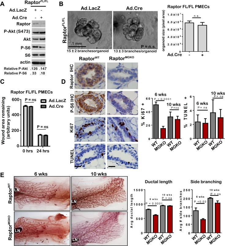
Fig 7. Unlike mTORC2, mTORC1 is dispensable for MEC survival and branching morphogenesis.
A-C. Raptor FL/FL PMECs and organoids were infected with Ad.Cre and Ad.LacZ, and cultured 10 days. A. Western analysis of PMECs cultured in the absence of serum. Quantitation was performed using Image J software and numbers represent P-Akt or P-S6 bands normalized to total Akt or S6 levels. B. Organoids were infected with Ad.Cre or Ad.LacZ photographed after 10 days in Matrigel culture. Representative images are shown. Average number of branches/organoid for each group is shown below the image. Average organoid size (pixels) ± S.D. is shown, Student’s T-test. N = 6 independent organoid isolates, analyzed in triplicate. C. PMECs from Raptor Fl/FL mice were infected with Ad.LacZ or Ad.Cre, grown to confluence and scratch-wounded. Monolayers were imaged. Total wound area remaining was measured 24 hours after wounding. Values shown are the avg ± S.D. N = 6 per time point, Student’s T-test. D-E. Mammary glands from virgin female RaptorWT and RaptorMGKO at 6 and 10 weeks of age were analyzed. N = 10 mice per genotype at each time point. Statistical analysis performed with Student’s T-test. D. IHC for Raptor, P-S6, Ki67+, and TUNEL+ nuclei in mammary glands of 6-week old mice. Representative images are shown. Scale bars = 50 microns. Average percent Ki67+ nuclei and TUNEL+ nuclei (± S.D) per total epithelial nuclei was determined E. Whole mount hematoxylin staining of mammary glands. Representative images are shown. LN = lymph node. The ductal length beyond the mammary lymph node was measured in whole mounted mammary glands. Average length (in microns) ± S.D. is shown. The number of T-shaped side branches was enumerated in whole mounted mammary glands. Values shown represent average number of side branches ± S.D.

