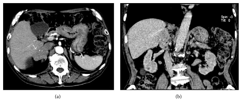
Figure 1.

(a) Computed tomography revealed an enhanced lobular tumor (white arrow) with calcification in the region of the right adrenal gland; (b) the border of the tumor at the liver and the right adrenal gland was not clear, and the tumor was close to the inferior vena cava (IVC) (white arrow).
