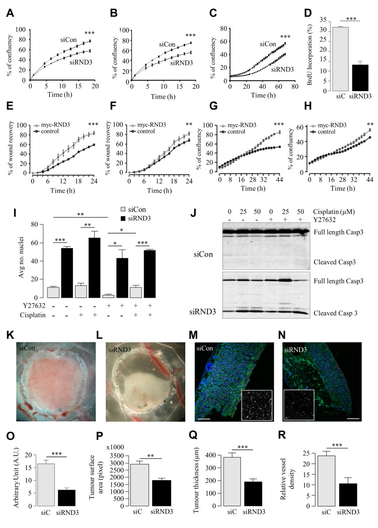
Fig 5. In vitro and in vivo RND3 silencing reduces cell proliferation and migration, induces apoptosis and reduces tumour mass.
A, B. (A) Invasion and (B) migration of U87 siRND3 and siControl cells determined by scratch wound assay. C. Proliferation of U87 siRND3 and siControl cells determined by confluence measurements. D. Percentage of cells showing BrdU incorporation into DNA in U87 siRND3 and siControl cells. E, F. Migration of RND3 overexpressing (E) 1321N1 and (F) T98G cells and Turbo-GFP control cells determined by scratch wound assay. G, H. Proliferation of RND3 overexpressing (G) 132N1 and (H) T98 cells. I. The average number of condensed nuclei in U87 siRND3 and siControl cells treated with Y27632 and/or cisplatin prior to imaging. J. Levels of cleaved Caspase 3 in U87 siRND3 and siControl cells with/without Y27632 and cisplatin treatment were determined by western blot. K, L. Representative image of 5 day old tumours derived from (K) wild-type and (L) U87 siRND3 cells implanted on the chicken CAM. M, N. Representative image of Ki-67 expression in 5 day old tumours derived from (M) wild-type and (N) U87 siRND3 cells. Magnification x10, scale bar 200μm, inset magnification x40. O-R. Quantification of (O) Ki-67 expression, (P) tumour surface area, measured as tumour area in pixels, (Q) tumour thickness, (R) relative density of blood vessels in 5 day old tumours. A-J. Data is representative of 3 independent experiments. K-R. Data is representative of 4 or more tumours. *** p < 0.001, ** p < 0.01, * p < 0.05, values +/- SEM.

