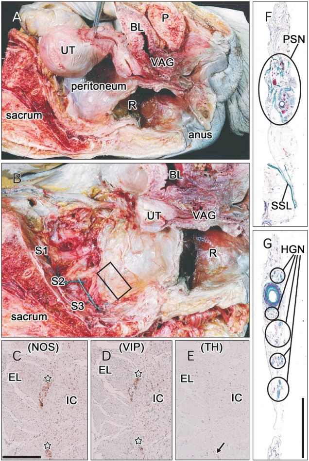
Fig. 1. Tissue sampling to prepare histological sections of the pelvic splanchnic nerve (PSN) from the left hemipelvis of a 78-year-old woman. (A) Picture of the body immediately after bisection of the pelvis. The hypogastric nerve (HGN) was sampled in front of the sacral promontory, and the body was cut transversely along the fifth lumbar vertebra. (B) Dissection of the sacral nerve roots (S1, S2, S3) during lateral traction of the pelvic viscera. The body of the uterus (UT) had been removed between steps shown in panels (A) and (B). The material analyzed, which included the third sacral root of the PSN, was obtained from the area indicated by the square in panel (B). (C-E) Positive controls for the present immunohistochemical assays: serial cross sections of the descending colon of the same specimen for the hypogastric and splanchnic nerves were incubated with antibodies against neuronal nitric oxide synthase (NOS) (C), vasoactive intestinal peptide (VIP) (D), and tyrosine hydroxylase (TH) (E). In contrast to the very restricted expression of TH (arrow in panel E), NOS and VIP were strongly and widely positive in the myenteric plexus (stars in panels C and D). (F, G) Masson trichrome staining of tissue strips containing the HGN (F) and PSN (G). Panels (C-E) are shown at the same magnification as are panel (F) and (G). Scale bars=0.5 mm (C), 10 mm (G). BL, bladder; EL, external longitudinal muscle layer of the colon; IC, internal circular muscle layer of the colon; P, pubis; R, rectum; SSL, sacrospinous ligament; VAG, vagina.

