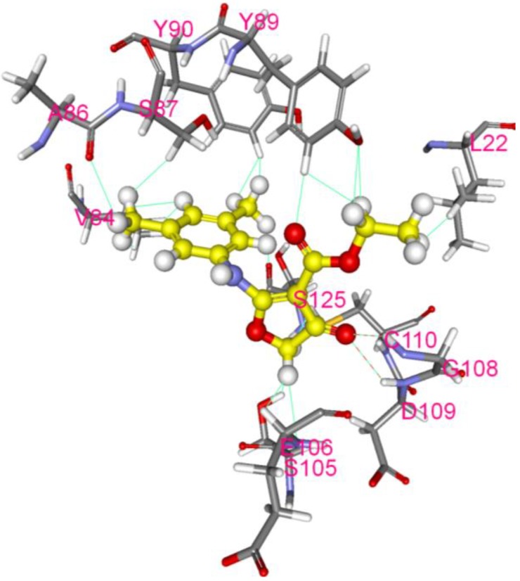
Figure 7.
Molecular modeling of interaction between CW-33 and 2A protease. Compound CW-33 (ball and stick, yellow) docked into the active site of EV-A71 2A protease sandwiched between N- and C-terminal domains. Compound CW-33 docked well with 3w95 via hydrogen bonding and hydrophobic reaction, binding amino acids shown as sticks and labeled.

