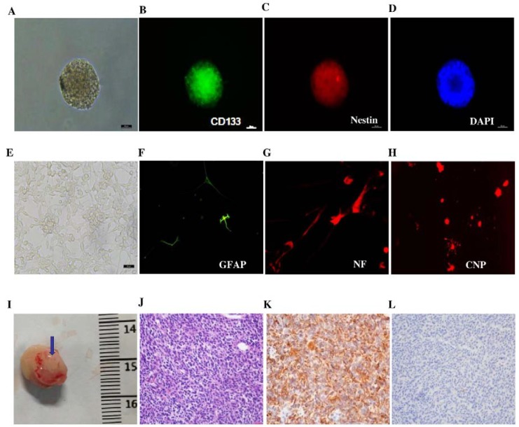
Figure 1.
Determination of GICs (412) in vitro and in vivo. (A) Suspended cells formed the self-renewing spheres in stem cell conditioned culture medium (100×); (B) Immunofluorescent staining of single suspended cells showed CD133-positive staining (200×); (C) Immunofluorescent staining of single suspended cells showed Nestin-positive staining (200×); (D) Immunofluorescent staining of DAPI (200×); (E) Morphology of serum-induced differentiation of GICs under a microscope (100×); (F) Immunofluorescent staining in differentiated GICs showed GFAP-positive staining (200×); (G) Immunofluorescent staining in differentiated GICs showed NF-positive staining (200×); (H) Immunofluorescent staining in differentiated GICs showed CNP-positive staining (200×); (I) The representative image of GIC tumorigenesis in NOD/SCID mice; the tumor is indicated by the blue arrow; (J) H & E staining showed the brain xenograft as GBM (200×); (K) Immunohistochemical staining in the xenograft section presented high expression levels of Nestin (200×); (L) Immunohistochemical staining of xenograft samples presented acicular expression of GFAP (200×).

