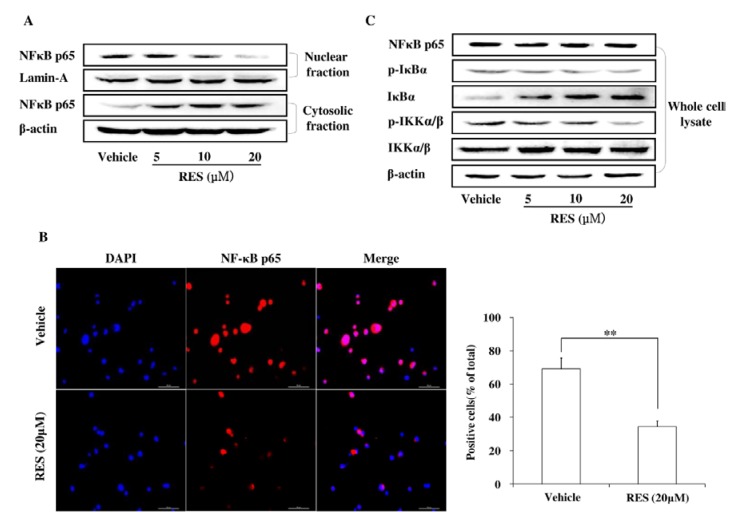
Figure 5.
RES suppresses GIC invasion through the inhibition of the NF-κB pathway. (A) GICs (412) were treated with DMSO (vehicle) and 5 μM, 10 μM, and 20 μM RES for 48 h, respectively. Cytoplasmic and nuclear fractions of GICs were isolated, and the concentration of nuclear and cytoplasmic protein was measured by western blotting with an anti-NF- kB p65 antibody. Lamin A and β-actin were used as loading controls; (B) GICs (412) were treated with DMSO (vehicle) or 20 μM RES for 48 h and then immunostained with anti-NF-κB p65 and DAPI. Then, randomly chosen fields were photographed (400×) and the number of positive cells was calculated. Data are shown as the mean ± SD of three independent experiments by Student’s t-test. * p < 0.05, ** p < 0.01, vs. Vehicle; (C) GICs (412) were treated with DMSO (vehicle) and 5 μM, 10 μM, and 20 μM RES for 48 h, and the expression of target proteins was detected by western blotting using anti-NF-κB P65, anti-IκBα, anti-p-IκBα, anti-p-IKKα/β, anti-IKKα/β antibodies. β-actin was used as a loading control.

