Figure 7.
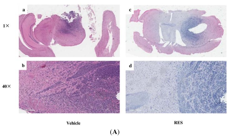
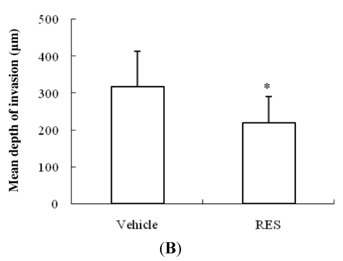
RES reduces invasion of GICs in vivo. 2 × 10⁵ GICs (412) in a volume of 5 μL PBS were stereotactically implanted into the brains of mice. Then, the mice were treated with propylene glycol (vehicle, 0.1 mL) or 10 mg·kg−1 RES (in 0.1 mL propylene glycol), respectively. (A) Representative results of H & E staining of GBM-bearing NOD/SCID mouse brain paraffin sections; (a) An H & E staining section of the brain from an NOD/SCID mouse treated with vehicle (1× original magnification); (b) a representative enlarged regional field (40× magnification) from (a); (c) an H & E staining section of the brain from an NOD/SCID mouse treated with RES (1× original magnification); (d) a representative enlarged regional field (40× magnification) from (c); (B) The depth of tumor cell invasion was evaluated as described in the “Materials and Methods” section. The bar graph represents the average depth of invasion (μm) at the edge of tumors derived from mice treated with vehicle or RES (n = 6/group). Data are shown as the mean ± SD. * p < 0.05, vs. Vehicle.


