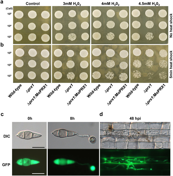
Figure 4. MoPRX1 complements S. cerevisiae PRX1 and localized in cytoplasm.
For yeast complementation test, cells were cultured overnight, diluted in phosphate-buffered saline at a final concentration adjusted to 2 × 10³ cells/μl, and aliquoted in 100-μl samples for each strain. Diluted 10 μl aliquots (10 μl containing 103, 104, and 105 cells/μl) of wild-type (BY4742), Δprx1 (YBL064C), and Δprx1:MoPRX1 were exposed to 3, 4 and 4.5 mM H2O2 on YPD agar plates after (a) with or (b) without 5-min heat shock at 50 °C. (c) Cellular localization of MoPRX1::GFP fusion protein in a conidium and appressoria of M. oryzae. (d) Infectious hypha expressing MoPRX1::GFP on rice sheaths at 48 hpi. DIC images were captured using a 20-ms exposure to transmitted light with a DIC filter. Fluorescence images were captured using a 400-ms exposure to absorbed light using a GFP filter. Bar = 10 μm.

