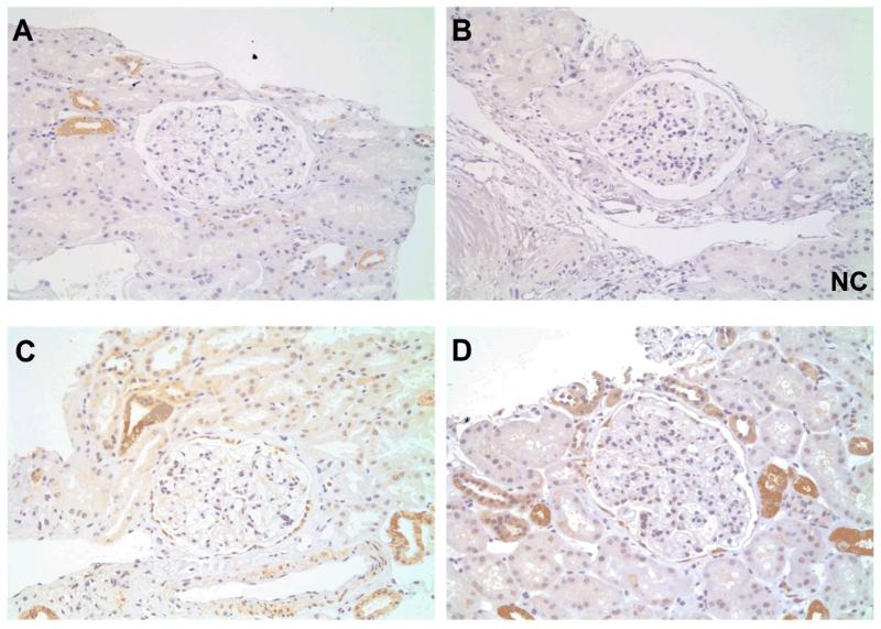
Figure 4. Total (T-) SYK expression non-proliferative glomerulonephritides.
(A & B) Sequential sections showing positive and negative control (NC) staining for T-SYK in thin basement membrane lesion. (C) T-SYK staining in minimal change disease. (D) T-SYK staining in idiopathic membranous nephropathy. In all cases, negligible glomerular staining is seen. There is intermittent staining of distal tubular epithelial cells, comparable to that seen in normal kidney tissue. All sections are immunoperoxidase stains with haematoxylin counterstain, x100-400 magnification. Negative control (NC) stains were performed by pre-incubating the primary antibody with the relevant immunising peptide.

