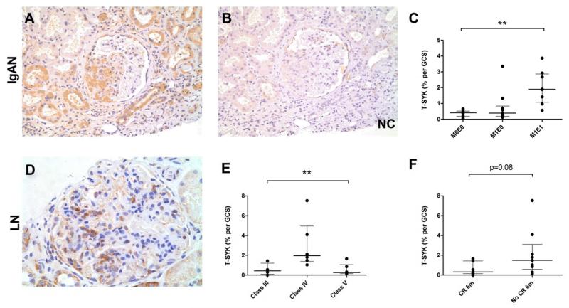
Figure 6. Total (T-) SYK expression in Immunoglobulin A nephropathy (IgAN) and lupus nephritis (LN).
(A & B) Sequential sections showing positive and negative control (NC) staining for T-SYK in IgAN, localised to a segmental area of endocapillary proliferation. (C) T-SYK detection in IgAN according to Oxford Class of disease; significant T-SYK detection was a feature of IgAN with endocapillary, and not mesangial, proliferation; M0E0, neither mesangial nor endocapillary proliferation; M1E0, mesangial proliferation without endocapillary proliferation; M1E1, both mesangial and endocapillary proliferation. (D) T-SYK detection in class IV lupus nephritis, localised to cells within the glomerular tuft. (E) T-SYK detection in LN according to ISN/RPS class of disease; SYK expression was a feature of class IV (diffuse proliferative) disease only. (F) T-SYK detection was higher in patients who failed to achieve complete remission (CR) at six months (6m) from initiating immunosuppressive treatment after this index renal biopsy. All sections are immunoperoxidase stains with haematoxylin counterstain, x200-400 magnification. Negative control (NC) stains were performed by pre-incubating the primary antibody with the relevant immunising peptide. GCS, glomerular cross section. ** p<0.01

