
Figure 1.
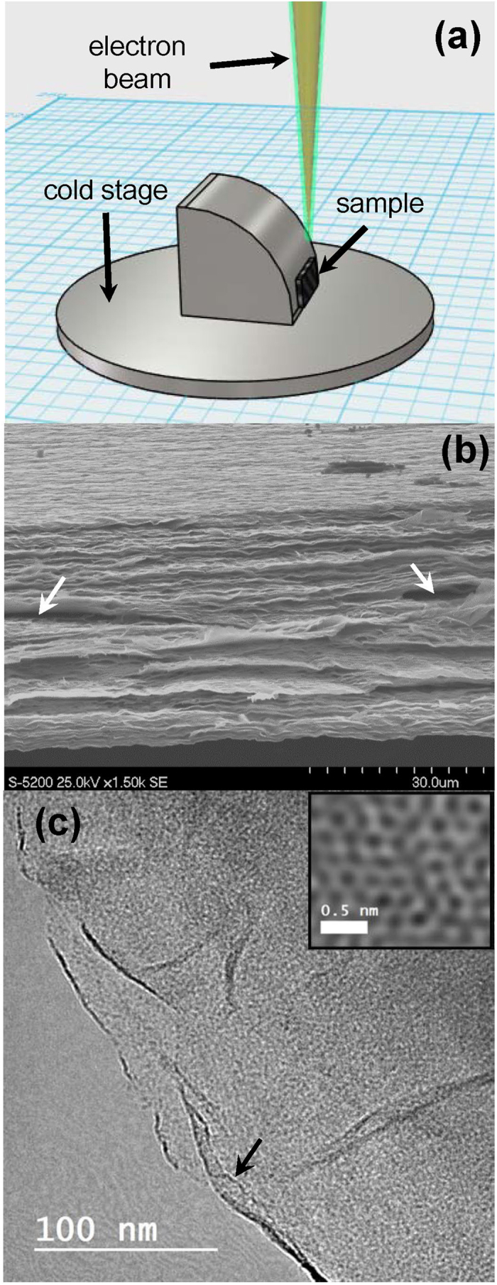
(a) Schematic image of the bespoke cold stage. The multilayer GO membrane is attached and the cross-section is exposed to the electron beam. (b) Photograph of the stage. (c) An SEM image of the multilayer GO membrane. (d) TEM images of a few-layer region of the multilayer GO membrane. The inset in (d) shows the structure at atomic resolution.