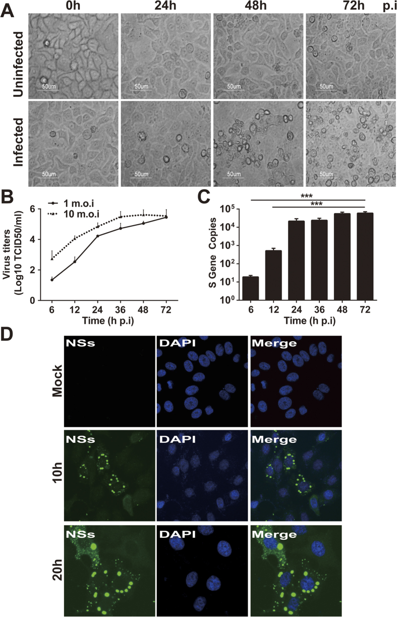
Figure 2. Human liver epithelial cells (HepG2) were susceptible to SFTSV infection.
HepG2 cells were mock-infected or infected with SFTSV at an m.o.i of 1. (A) Significant CPE was observed in infected cells (magnification 400×) at 48 and 72 hrs p.i. (B) Replicative curve of SFTSV in infected HepG2 cells. Culture media of the infected HepG2 cells were taken at various time points p.i., ten-fold serially diluted, and inoculated in Vero cells in 96-well plates. Infectious virus titers were determined after the cells were fixed and stained with an HRP-conjugated anti-viral NP antibody, followed by colorimetric development with TMB substrate. Infection foci were counted and TCID50 calculated based on the Reed and Muench method. (C) Quantitation of viral RNA in infected HepG2 cells. Total RNA was prepared from uninfected and infected cells at 6, 12, 24, 36, 48, and 72 hrs p.i. and viral RNA were qualified with real-time RT-PCR after reverse transcription. Specific primers for the S gene were used to quantify copy numbers of the S gene. The experiments were repeated at least three times and the data from one representative with two repeats were presented (* p < 0.05). (D) SFTSV antigens detected in infected HepG2 cells. Both infected and control cells were fixed at 10 or 20 hrs p.i. After permeablization with PFA, the cells were incubated with rabbit anti-NSs antibody at a dilution of 1:100, followed by staining with Alex Fluro488-conjugated secondary antibody.

