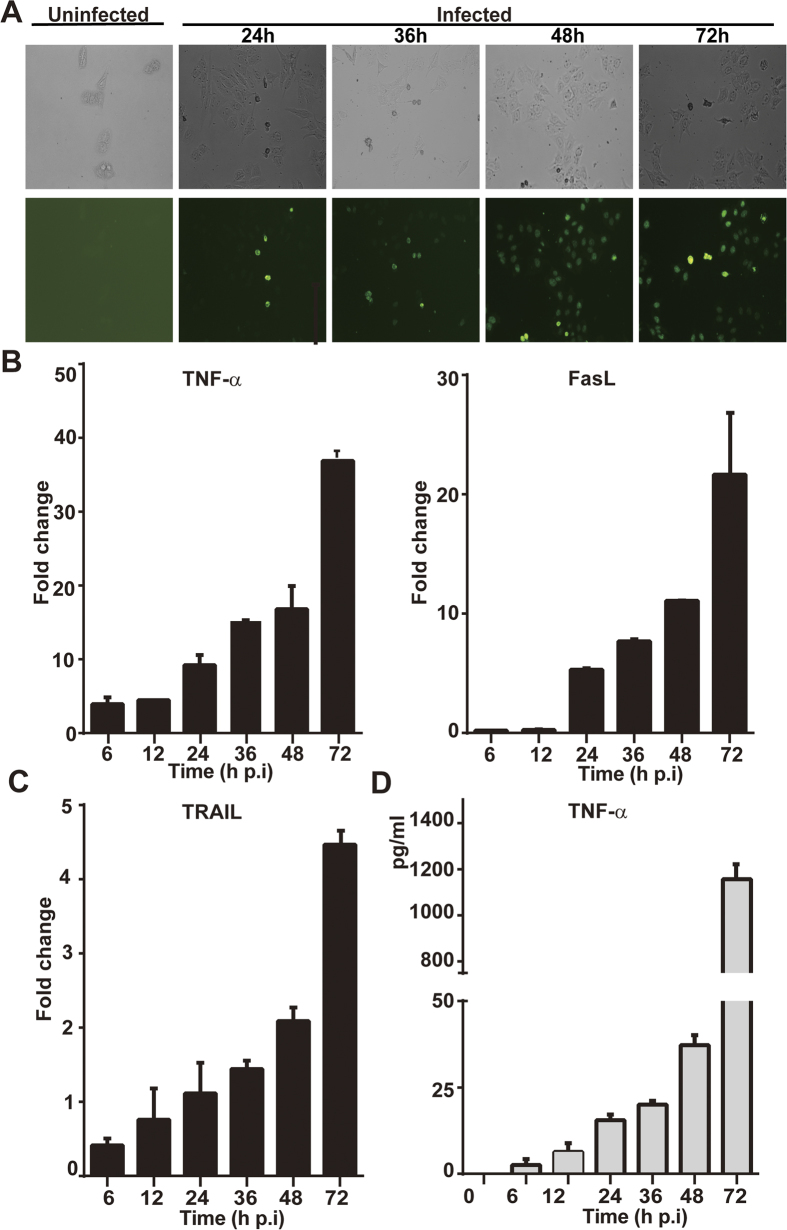
Figure 3. Apoptosis induced by infection of SFTSV in HepG2 cells.
(A) Cells were infected with SFTSV at an m.o.i of 1. In situ apoptosis was detected from 24–72 hrs p.i. by an FITC-dUTP labeled TUNEL assay. (B) Transcript levels of TNF-α, FasL, and TRAIL from 6–72 hrs p.i. in infected cells were measured by realtime RT-PCR with specific primers to corresponding cytokines. (C) Concentration of TNF-α induced in infected cells. Cultural media of cells infected with SFTSV were collected at various times points p.i. and used for ELISA measurement of the concentration of TNF-α. The assays were repeated at least three times.

